Owner Fact Files: Elbow Dysplasia
- Nancy Veterinary Physiotherapist

- Jan 28, 2021
- 6 min read
The 6th instalment in the Owner Fact Files - Elbow Dysplasia, sometimes better known as 'development elbow disease', the most common cause of forelimb lameness seen in dogs.
Elbow Dysplasia is used as an umbrella term for a range of complex disorders of the elbow - read on to find out more about each one.
What is Elbow Dysplasia?

Elbow dysplasia (ED) is abnormal development of the elbow joint – if the three bones that make up the joint don’t fit perfectly, the abnormal development occurs. The condition is slightly different from Hip Dysplasia (HD), as with HD the hips develop abnormally causing arthritis to develop. With ED, the elbows develop abnormally but this causes additional issues within the joint, alongside arthritis.
To understand the condition, you should have good knowledge of how a healthy elbow should look and function. The elbow works as a hinge joint, with 3 bones coming together - the humerus, radius and ulna. When these 3 bones fit together perfectly, easy and smooth movement occurs at the joint.

Unfortunately in some cases there is an abnormal fit at the joint, read on to find out why this happens and the impact it has on the dog.
What causes Elbow Dysplasia?
Elbow Dysplasia is NOT congenital, meaning dogs that have it are born with normal elbows. As they begin to grow, the abnormalities start to develop causing the joint to have a poor fit. If a joint does not fit together correctly, stress is put on the bones which leads to damage and loss on function.
There are a few things that can cause poor fit of the elbow:
1. In some cases the radius and ulna bone grow at different rates so one of the bones ends up longer than the other. This causes a severe overload of force on the ulna bone, which damages it over time.
2. The radius bone repetitively hits the ulna, causing small fractures.
3. The ulna is the wrong shape, so does not fit against the humerus. The bones will rub against each other causing the cartilage on the end of the humerus to erode.
There are various factors that can influence the development of ED in dogs, starting with Genetics. Every dog with ED will have the genes for the condition, it cannot be caused solely by environmental factors, such as poor diet. ED is an inherited condition, which is why dogs known to have it should not be used for breeding. There is a system in place to score dogs' elbows from X-Rays to try and reduce the cases of ED. The condition is more commonly seen in large breed dogs such as the Bernese Mountain Dog, Labrador, Golden Retriever, and German Shepherd, and it affects both elbows in 80% of cases.
There are environmental factors, that along with the genes, can have an impact on the development of ED. Diet and body weight are described as risk factors for elbow conditions, with excess weight putting excess strain on the joint. Poor diet can also influence growth which could alter the development of the joint. Certain types of exercise may also increase the risk of ED - chasing a ball/stick, due to the concussive forces on the joints and the twisting and turning.

What is happening inside the elbow joint? (the science bit)
So as previously mentioned, Elbow Dysplasia, is an umbrella term for several different conditions of the elbow, these are:
Fragmented coronoid process (FCP)
Ununited anconeal process (UAP)
Osteochondritis dissecans (OCD)
Medial compartment disease
Sometimes more than one of these problems can occur in the same joint. So let's discuss each of these conditions in a bit more detail.
Fragmented coronoid process (FCP)
On the ulna bone there are 2 small bony protrusions called coronoid processes, and in some elbows, these parts of the bone have a developmental defect. One of the coronoid processes develops a crack and separates from the rest of the bone. This causes pain and instability in the elbow.

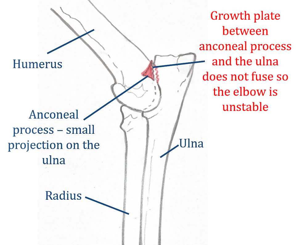
Ununited anconeal process (UAP)
The anconeal process is a small projection of bone also on the ulna, which forms part of the back surface of the elbow. There is a growth plate between the anconeal process and the rest of the ulna that should fuse in normal development at around 5 months of age. In cases of UAP, the growth plate does not fuse making the elbow unstable. In some cases the loose fragment of bone will float around in the joint causing more pain.
Osteochondritis dissecans (OCD)
OCD is the most common of the elbow conditions and describes abnormal development of cartilage on the end of a bone, in the case of ED on the bottom of the humerus. The cartilage lifts off, away from the bone, creating a loose cartilage flap. Small parts of cartilage can also break off and float around in the joint space. Without the protection of the cartilage, the development of the bone underneath is affected and osteoarthritis develops.

Medial compartment disease
This condition is similar to OCD, as the cartilage wears away on the inside or medial side of the elbow joint. This change causes severe arthritic changes, and has the poorest prognosis of all of the conditions.
Symptoms
You may start to see signs of ED in your dog from 6 months old, with diagnosis commonly happening between the age of 6-12 months. In some cases ED will not be discovered until the secondary arthritis has become advanced, and the symptoms of this will draw attention to the underlying issue.
The signs that you may notice are;
- Forelimb lameness - worse after exercise and typically doesn't resolve after rest
- Stiffness
- Reluctance to exercise
- Difficulty coming down stairs
- Front feet appear turned out
- Pain/swelling/heat at the joint
- 'Paddling' or 'flipping' of front feet when walking
If you see any of these symptoms in your dog and you have concerns please speak to your vet. ED can vary in severity, so you may not see all of these signs.
Treatment
The treatment selected will depend on the specific condition that affects your dog's elbows. Conservative management may be appropriate in some cases, where the symptoms are milder and the disease has less impact on your dog's life. Things that can be done to manage ED include body weight management, pain relief medication and changes to exercise routine. Additional physiotherapy/hydrotherapy also helps to support the elbow by building muscle and maintaining range of motion and functionality.
For dogs that require surgery, there are a few options available. If there are any loose fragments of bone or cartilage in the joint these will be removed by an Arthroscopy. In cases that are caused by a mismatch of length between the radius/ulna, the longer bone can be cut to create a better fit at the joint. This procedure is called a Proximal ulnar osteotomy (PUO).
If the cause of abnormal development is the ulna and radius colliding, surgery can be used to release part of the biceps tendon from the surface of the ulna. This is called a Biceps ulnar release (BURP) and neutralises the forces to stop the bones from hitting each other. In cases of Medial compartment disease, a Sliding humeral osteotomy can be performed to cut the humerus and fix it in a new place. This takes the pressure away from the diseased side of the joint.
Finally, if the osteoarthritis in the elbow is severe, with little to no cartilage left, a Total elbow replacement can be done. This would be a last resort and is a very specialist surgery only available at a few practices in the UK.
How can physiotherapy help?
Physiotherapy is a very useful tool in the treatment of ED, whether you are using conservative management or in rehab following surgery. Pain management is the priority, whether this be pain in the joint or pain in other muscles/joints that have been working harder to compensate for the diseased elbows. A physiotherapist can also advise on a safe return to exercise where we choose targeted exercises to help build muscle in the forelimbs to support the elbows. Certain exercises will also be used to encourage your dog to use their elbow, keep it flexible and increase range of motion.
All of the techniques used can help manage the secondary osteoarthritis in the elbow, which will inevitably develop with ED. Ongoing sessions with a physio help to keep on top of this, and potentially slow the progression.

For more information on how physiotherapy could benefit your dog with Elbow Dysplasia please get in touch - vetphysionancy@outlook.com // 07795163445.
Prognosis
The sooner elbow problems are detected the better the outcome will be. Unfortunately in all cases, osteoarthritis will develop in the joint, but if dealt with quickly this can be slowed down so the elbow can stay functional for longer. Due to the arthritis, dogs with ED will need lifelong care - physiotherapy maintenance programmes can be helpful to monitor your dog and keep them on track.
If you have any more questions about this condition feel free to contact me! Remember to come back next month for the next Owner Fact File, and leave me some feedback if you enjoyed this blog.
If there is anything I can do to help your animals please drop me a message - Nancy
M: 07795163445
Find me on social media - Facebook and Instagram @vetphysionancy


Comments